|
|
|
News |
|
|
| |
|
| |
 |
| |
|
| |
| |
|
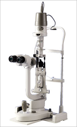
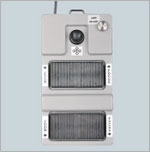
Operating Microscope Posterior Segment |
|
|

|
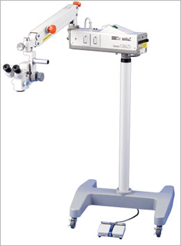
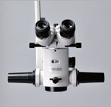
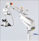
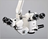
Operating Microscope OM-18
|
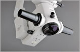
Compact Microscope
The suspension arm has been moved to the
side of the microscope, opening up the
operating surgeon’s front field of view.
-
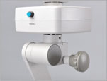
The light source is placed away from the
microscope section. Cold light coaxial
illumination via fiber light guide is
used. The fiber light-guide illumination
system is particularly low in heat
transmission, and reduces the projection
of the filament image as hot spots,
achieving excellence in safety. By
separating the light source from the
microscope section, we have not only
successfully removed the affect of the
heat generated by the light source, but
also achieved a compact body
-
The motorized zoom magnification changer
used in the OM-18 Operating Microscope,
with its diverse magnification ranging
from 4.6x to 27.4x, guarantees a
strain-free operating environment
-
The newly designed illumination optics
provides the φ55mm illumination field
with consistent brightness throughout,
from low magnification to high
magnification
-
The newly designed tilting mechanism
allows for smooth angle adjustment. It
is effective in adjusting the
irradiation angle of the illumination by
changing the angle of the microscope
towards the operated eye.
|
 |
| |
|
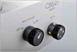
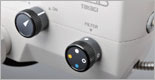
Diverse illumination optics
With its diverse illumination optics,
suitable lighting for the operating
environment can be selected.
Equipped with the Red Reflex
Illumination IN/OUT Switching Mechanism
The red reflex is enhanced with the use
of the red reflex illumination mechanism
that is built into the internal part of
the microscope. When this mechanism is
used in combination with 6°
illumination, an even more comfortable
illumination for cataract surgeries is
achievable. Furthermore, depending on
the eccentricity of the operated eye,
adjustment in the range from +2°to -2°
can be made, which further improves the
illumination environment.
Equipped with the 6° Illumination IN/OUT
Mechanism
By using 6° coaxial illumination, which
is also called the stereoscopic
illumination, shades that correspond to
the convex/concave of the observation
field are formed, providing a
three-dimensional impression. By
adjusting the brightness with the Fade
In/Out function in the OM-18, the red
reflex becomes easier to see during a
cataract surgery.
|
 |
| |
|
Protection against phototoxicity
During a cataract surgery, light that
used to be absorbed by the crystalline
lens would reach the retina after
removal of the crystalline lens,
inducing retinal phototoxicity. The
OM-18 has built-in filters that shield
photodamage-causing wavelengths to
protect the patient’s retina against
phototoxicity.
UV Filter(Permanent feature built-in)
In order to shield the wavelengths of
harmful ultraviolet rays that cause
retinal damage, the OM-18 has a built-in
UV filter, protecting the patient’s eye
against the harmful ultraviolet rays.
Blue-Cut Filter
The blue-cut filter shields visible blue
wavelengths in order to protect the
patient’s eye against the blue-light
hazard. This filter can be selected In
or Out during an operation. (Also called
the yellow filter)
Heat-Absorbing Filter(Permanent feature
built-in)
The OM-18 has a built-in heat-absorbing
filter that shields the transmissive
infrared rays that affect the retina and
the choroid, thus protecting the
patient’s eye.
Retina Shield Filter
The retina shield filter is built-in in
order to protect the retina, after
crystalline lens’ removal, against the
intense illumination. This filter can be
selected In or Out during an operation. |
 |
| |
|
Newly designed apochromatic objective
In the optics of the microscope, light
that has been transmitted through the
prism is dispersed into seven colors.
This dispersion causes chromatic halos
and fringing in images as well as
blurred images. Apochromatic correction
corrects the wavelengths that cause such
aberrations.
With the apochromatic correction, the
OM-18 has reduced chromatic halos and
fringing, further improving resolution
and contrast. The correction given to
indigo (G-line, 435nm), in particular,
contributes to clearer images. |
 |
| |
|
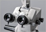
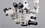
Surgeon-friendly tiltable binocular
tubes with converging optics
Visual axes are slanted inward by 6°,
which enables easy fusion at a natural
eye position. Being equipped with a
depth-adjusting diaphragm as a standard
feature, fine tuning of the microscope’s
depth range can be easily preformed.
-
High-eyepoint eyepieces have enabled a
wide visual field. For optics,
multi-coated lenses of high grade are
used, delivering crisp bright images.
-
The angle adjustment is extensive, from
upright (straight) to 60° (inclined)
depending on the posture and the
physique of the operating surgeon,
ensuring the optimum position.
|
 |
| |
|
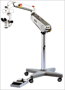
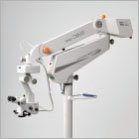
Improved vibration damping
The counter-balanced arm, a new
mechanism, has reduced shaking in the
head caused when the arm is moved, to a
minimum, particularly in the X-Y
direction.
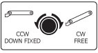
Safety stopper mechanism
The OM-18 is equipped with a safety
stopper mechanism, where the lowest arm
position can be easily set according to
the height of the operating table. |

 |
| |
|
Improved transportability and excellent
arm storage
Ease of transportation and secure lock
Large-diameter wheels and a maneuvering
handle have further imporoved its
transportability. The OM-18 can be moved
across different floor levels with ease.
Wheel locks secure the operating
microscope when it needs to be fixed
after transportation. |
 |
| |
|
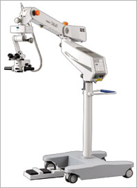
Excellent storage
By folding the counter-balanced arm, the
OM-18 can be stored in a limited space. |
 |
| |
|
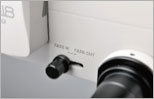
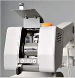
Lamp switching at the touch of a lever
With the use of the fiber light-guide
system, the dual lamp source is
positioned away from the microscope
section, allowing lamps to be switched,
outside the sterile field, at the touch
of a lever even during an operation.
Lamp Condition indicators on the control
panel show the status of the halogen
bulbs stored in the light source
section, which can easily be checked
prior to an operation. |
 |
| |
|
Foot controller
The foot controller allows 8-way
operations: Main lamp on, Main lamp off,
X direction movement, Y direction
movement, Focusing up, Focusing down,
Zooming up and Zooming down. There is
also a water-resistant version of the
foot controller where the layout of the
focus and zoom pedals can be selected to
suit user’s performance. |
 |
| |
|
|
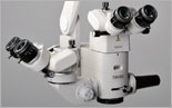
Diverse options
Coaxial stereoscopic microsope for
assistant
The OM-18 provides the assistant with a
stereoscopic view in the optical path
coaxial to that of the operationg
surgeon. Magnification can be changed
manually in three steps. The assistant’s
microscope can be mounted either side of
the operating surgeon’s microscope.
Visual axes of the binocular tubes are
slanted inward by 6°, which allows easy
fusion at a natural eye position. Being
equipped with a depth-adjusting
diaphragm as a standard feature, fine
tuning of the microscope’s depth range
can be easily performed. The angle of
the binocular tubes is adjustable
extensively from upright (straight) to
60°
(inclined) depending on the posture and
the physique of the assistant, ensuring
the optimum position.
Eyepieces:12.5x
Objective:F=175
Total magnifications:4.1x, 6.8x, 11.4x
Real fields of view:φ54.7, φ32.8, φ19.7 |
 |
| |
|
|
Beam splitter / TV Camera adaptor
The beam splitter and the camera adaptor
are required to install a C-mount CCD
camera. The beam splitter is equipped
with an In/Out switching lever.
The camera adaptor is equipped with a
diaphragm mechanism. Images taken during
an operation can be recorded in the
peripheral equipment connected.
Beam splitter to be used
O11-02(40% to the operating surgeon:60%
to the camera)
O11-03(80% to the operating surgeon:20%
to the camera)
TV camera adapter to be used
O08-11 F=50mm
O08-05 F=90mm |
 |
| |
|
Video beam splitter
The single-chip CCD camera module(1/4”,
400,000 pixels, Y/C, composite output)
and the control unit are built into a
single body, achieving a compact and
low-priced adapter. The power source and
signal are transmitted on a single
cable. Two (2) types of the Video Beam
Splitters are available.
Video beam splitter to be used
O08-15(NTSC)
O08-16(PAL) |
 |
|
Oculus SDI/BIOM 3 and 4 adaptations are
available for OM-18 |
|
Specifications
|
Microscope |
Magnification changer |
Motorized zoom type (zoom ratio 1:6) |
|
Objective |
F=175mm (apochromatic corrected optics) |
|
Eyepieces |
12.5x(high-eyepoint & wide field) |
|
Binoculars |
Tiltable binoculars with converging
optics F=160mm |
|
Total magnifications |
4.6x ~ 27.4x |
|
Real fields of view |
φ49.2mm to φ8.2mm |
|
Focusing stroke |
50mm (with centering function) |
|
X-Y movement stroke |
±25mm in each direction(with centering
function) |
|
Illumination |
System |
Cold light coaxial illumination by fiber
light guide |
|
Light source |
15V 150W halogen lamp |
|
Field of illumination |
φ55mm |
|
Field of red reflex illumination |
φ15mm |
|
Illumination control |
Continuous adjustment |
|
Filters |
Heat-absorbing, UV(permanent feature
built-in), Blue-cut Cobalt Blue, Retina
Shield(for protection of the retina) |
|
Arm,base |
Mount |
Floor stand |
|
Maximum arm extension |
1260mm |
|
Arm vertical stroke |
500mm |
|
Base size |
700mm × 740mm |
|
Foot controller size |
310mm × 200mm× 105mm |
|
Others |
Weight |
160kg |
|
Power consumption |
400VA |
|
Power supply |
AC120V, AC230V ; 50/60Hz |
|
|
|
You can Also Browse other Products of
Tagaki |
|
|
| |
|
|
| |
|
| . |
|