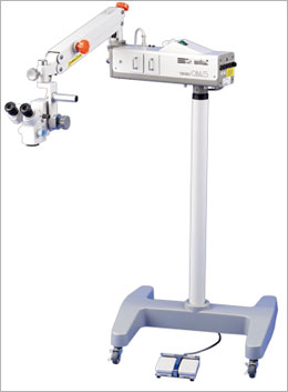
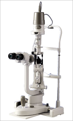
Slit Lamp Microscopes - SM-70N
Slitlamp
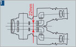
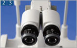
22mm Inter-optical Path Distance
As a general rule, the longer the
distance between the two optical paths,
the better the stereoscopic view, but
the narrower the binocular field of view
in fundus examination. In reverse, the
shorter the distance between the optical
paths, the poorer the stereoscopic view,
but the wider the binocular field of
view in fundus examination.
With this in mind, we have achieved
optics suitable for fundus examination
by choosing the optimal 22mm as the
inter-optical path distance. |
|
| |
|
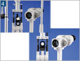
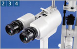
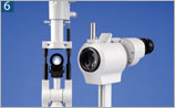
Converging Binocular Tubes
Binocular tubes with a 6-degree
convergence provide easy binocular
fusion, ensuring stress-free
observation.
Advanced multi-coating is applied to all
lenses used in the microscope for
excellent optical performance; bright
images free from flare and ghost are
obtained, improving the quality of
examination and treatment. |
|
| |
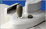
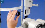
Helicoid Mechanism
As the diopter adjustment rings do not
rotate with the eye caps, the selected
diopters will not be accidentally
changed during use.
In addition, the 16x high-eyepoint
wide-field eyepieces enable observation
over a wider field. |
| |
|
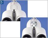
Specially-coated Mirror and Diffuser
The reflecting mirror has been given a
special coating, effectively reducing
harmful infrared and ultraviolet rays to
protect the eye being observed against
phototoxicity while providing an
exceptionally natural view in the
visible light range.
When photographing the anterior segment
of the eye, the diffuser, a standard
feature, can be used to extensively
illuminate the region being observed. |
|
| |
|
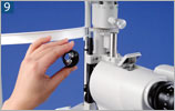
Integrated Control
The joystick for XYZ movement, its top
button for the light booster function
(which also serves as a trigger button
for capturing images when connected to
an imaging device), and the rheostat
adjacent to the joystick for light
intensity adjustment can all be
controlled with one hand. This ensures a
smooth and swift
examination.
Furthermore, the joystick mechanism
provides outstanding control from coarse
to fine movement of the slitlamp base. |
|
| |
|
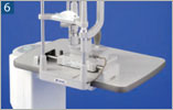
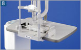
Slitlamp with Integrated Base
By integrating it with the base, the
sturdiness of the chin rest assembly has
improved dramatically. Now that the base
is integrated, there is no need to be
selective with the shape of fittings for
the chin rest assembly or its
installation method. The slitlamp can
now be set up very easily on any type of
instrument table. |
|
| |
|
Headrest/Finger Rest
The headrest serves not only as a
patient's headrest but also as a finger
rest for the examiner holding an
indirect lens upon fundus examination.
The finger rest feature is designed such
that the examiner can hold the indirect
lens steady. It also reduces arm fatigue
from lengthy fundus examination. |
|
| |
|
Right Eye / Left Eye Recognition
Sensor and Signal Output Function
The right eye/left eye recognition
sensor is now built-in so that the
slitlamp works well with an image filing
system. Right eye/left eye recognition
signal is output once the slitlamp is
aligned to the eye to be tested.
The cable-end connector of the
connecting cable (optional) varies
according to the image filing system
used. Contact our Sales Department for
details. |
|
Major Specifications
|
Microscope |
Type |
Galilean converging binocular
stereomicroscope |
|
Magnification changer |
Five position rotating drum |
|
Eyepieces |
16x wide-field, high-eyepoint |
|
Total magnifications |
6.3x, 10x, 16x, 25x, 40x |
|
Real field of view |
Ø35.9, Ø23.3, Ø14, Ø8.8, Ø5.5mm |
|
Interpupillary adjustment |
53mm〜84mm |
|
Diopter adjustment range |
+/-7diopters |
|
Cross-Slide Base |
Longitudinal (coarse) movement |
90mm |
|
Lateral (coarse) movement |
110mm |
|
Horizontal (fine) movement |
15mm |
|
Vertical movement |
+/-15mm |
|
Chinrest Unit |
Elevation stroke |
70mm |
|
Fixation light source |
Red LED |
|
Illumination Unit |
Slit width |
0-10mm continuously variable (at 10mm,
slit becomes a circle) |
|
Slit length |
1-10mm continuously variable |
|
Aperture diaphragm |
Ø10, Ø5, Ø3, Ø2, Ø1, Ø0.2mm |
|
Filters |
HA (heat-absorbing), G (red-free), B
(excitation), UV (Ultraviolet radiation
cut) |
|
Light source |
12V 30W halogen bulb |
|
Power Unit |
Input voltage |
AC100V-230V |
|
Maximum power consumption |
64VA |
|
Dimensions & Weight |
Base dimensions |
359mm(W) x 328mm(D) |
|
Weight |
13.3kg |
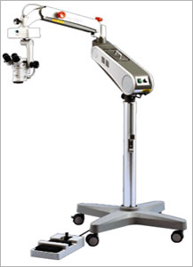
Slitlamp Microscope SM-90N
22mm Inter-optical Path Distance
As a general rule, the longer the
distance between the two optical paths,
the better the stereoscopic view, but
the narrower the binocular field of view
in fundus examination. In reverse, the
shorter the distance between the optical
paths, the poorer the stereoscopic view,
but the wider the binocular field of
view in fundus examination.
With this in mind, we have achieved
optics suitable for fundus examination
by choosing the optimal 22mm as the
inter-optical path distance. |
|
| |
|
Converging Binocular Tubes
Binocular tubes with a 6-degree
convergence provide easy binocular
fusion, ensuring stress-free
observation.
Advanced multi-coating is applied to all
lenses used in the microscope for
excellent optical performance; bright
images free from flare and ghost are
obtained, improving the quality of
examination and treatment. |
|
| |
Helicoid Mechanism
As the diopter adjustment rings do not
rotate with the eye caps, the selected
diopters will not be accidentally
changed during use.
In addition, the 12.5x high-eyepoint
wide-field eyepieces enable observation
over a wider field. |
| |
|
Motorized zoom system
The TAKAGI slitlamp technology is
combined with an electric zoom mechanism
unique in its class. The zoom mechanism
allows magnification to be changed over
a range of 5.5x-32x to provide optimum
magnification in clinical applications. |
| |
|
Magnification display using one-touch
flip-up mirror
The current magnification is displayed
using the one-touch flip-up mirror, thus
allowing photography at the fixed
magnification when taking multiple
images. Interior illumination of the
display unit ensure that the
magnification display is visible even in
dark surrounding.
Fitting the combination adapter (S10-17)
ensures that the magnification is
displayed even when using an imaging
system. |
|
| |
|
|
Specially-coated Mirror and Diffuser
The reflecting mirror has been given a
special coating, effectively reducing
harmful infrared and ultraviolet rays to
protect the eye being observed against
phototoxicity while providing an
exceptionally natural view in the
visible light range.
When photographing the anterior segment
of the eye, the diffuser, a standard
feature, can be used to extensively
illuminate the region being observed. |
|
| |
|
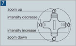
Centralized control system
In addition to the ability to move the
slitlamp, and 3D movement in the X,Y,
and Z directions by joystick the
provision of a trigger button (also
functions as the light boost button) at
the top,
and connection to video equipment allows
the examiner to acquire excellent images
while looking through the slitlamp. The
newly developed X-Y control button
fitted for the first time to the
slitlamp allows the zoom up-down and the
intensity increase and decrease to be
controlled with one hand. The X-Y
control button may be rotated 90
degrees, thus allowing the examiner to
change the direction as necessary.
* Light boost and trigger do not work
simultaneously |
|
| |
|
Slitlamp with Integrated Base
By integrating it with the base, the
sturdiness of the chin rest assembly has
improved dramatically. Now that the base
is integrated, there is no need to be
selective with the shape of fittings for
the chin rest assembly or its
installation method. The slitlamp can
now be set up very easily on any type of
instrument table. |
 |
|
|
|
Headrest/Finger
Rest
The headrest serves not only as a
patient's headrest but also as a finger
rest for the examiner holding an
indirect lens upon fundus examination.
The finger rest feature is designed such
that the examiner can hold the indirect
lens steady. It also reduces arm fatigue
from lengthy fundus examination. |
 |
|
|
|
Right Eye / Left Eye Recognition
Sensor and Signal Output Function
The right eye/left eye recognition
sensor is now built-in so that the
slitlamp works well with an image filing
system. Right eye/left eye recognition
signal is output once the slitlamp is
aligned to the eye to be tested.
The cable-end connector of the
connecting cable (optional) varies
according to the image filing system
used. Contact our Sales Department for
details. |
|
|
Microscope |
Type |
Galilean converging binocular
stereomicroscope |
|
Magnification changer |
motorized zoom |
|
Eyepieces |
12.5x wide-field, high-eyepoint |
|
Total magnifications |
5.5x - 32x |
|
Real fields of view |
φ 40.9 - φ 6.8mm |
|
Interpupillary adjustment |
53mm - 84mm |
|
Diopter adjustment range |
+/-5diopters |
|
Cross-Slide Base |
Longitudinal (coarse) movement |
90mm |
|
Lateral (coarse) movement |
110mm |
|
Horizontal (fine) movement |
15mm |
|
Vertical movement |
+/-15mm |
|
Chinrest Unit |
Elevation stroke |
70mm |
|
Fixation light source |
Red LED |
|
Illumination Unit |
Slit width |
0-10mm continuously variable (at 10mm,
slit becomes a circle) |
|
Slit length |
1-10mm continuously variable |
|
Aperture diaphragms |
φ 10, φ 5, φ 3, φ 2, φ 1, φ 0.2mm |
|
Filters |
HA (heat-absorbing), G (red-free), B
(excitation), UV (Ultraviolet radiation
cut) |
|
Light Source |
12V 30W halogen bulb |
|
Power Unit |
Input voltage |
AC100V - AC230V |
|
Maximum power consumption |
64VA |
|
Dimensions & Weight |
Base dimensions |
359mmW×328mmD |
|
Weight |
13.5kg |
|